BIGGER PICTURE Scientific discovery
The one-percent protein assembly that an average could hide
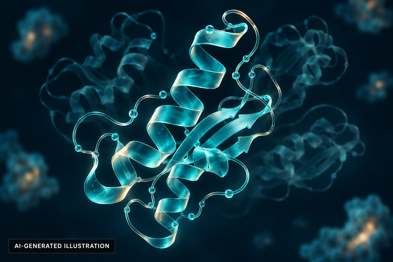
Proteins move and assemble in different ways. CryoDRGN helped scientists recover that variety from microscope images—including a rare ribosome structure that did not follow the expected assembly order.
- Surprising observation
- A rare assembly state in about 1% of ribosomes
- Starting material
- Experimental 2D cryo-EM images
- Result
- Multiple 3D shapes from one dataset
How the idea works
Collect frozen molecular views
Cryo-EM supplies experimental images from many particles.
Separate structural variation
A neural model represents a continuum of 3D states.
Inspect rare assemblies
Biologists examine minority states and test their interpretation.
A cell’s machine takes an unexpected route
Ribosomes build proteins, and assembling a ribosome is itself a complex construction project. Using cryoDRGN, MIT researchers found a small population in which a component normally added late appeared before the usual foundation was complete. It was present in roughly one percent of the examined ribosomes—a detail that a dominant average structure could obscure.
Sources: [1]
Frozen snapshots do not all show the same shape
Cryo-electron microscopy records many noisy two-dimensional views of molecules frozen in ice. Conventional reconstruction works especially well when particles share a stable structure. The challenge grows when a sample contains parts moving through different positions or complexes at different assembly stages. Treating all of those images as one object can discard precisely the variation scientists want to understand.
Learn a landscape of molecular states
CryoDRGN uses a neural representation to reconstruct a distribution of three-dimensional density maps. Researchers can inspect clusters, generate representative structures and explore paths between shapes. The published work recovered variation in ribosome and RAG-complex datasets and large flexible changes in a spliceosome. This uses measured images, rather than predicting a protein from its sequence alone.
A better scientific lens
The immediate benefit is richer structural evidence for biologists, supported by openly released software. It is not a filmed sequence of one molecule moving, nor a demonstrated new medicine. The inspiring principle is to search an existing dataset for meaningful minority states instead of forcing every observation into a single neat average.
Sources: [2]
The original sources
- MIT NewsMachine-learning model helps determine protein structures
- Zhong and colleagues / MIT repositoryCryoDRGN: reconstruction of heterogeneous cryo-EM structures using neural networks
- Zhong and colleagues, ICLRReconstructing continuous distributions of 3D protein structure from cryo-EM images